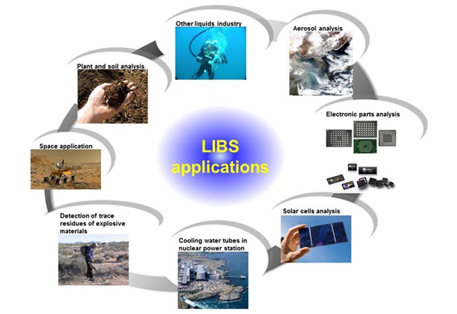
Laser induced breakdown spectroscopy (LIBS) has been recognized as a powerful and versatile technique for materials analysis, especially for the applications in which real-time analysis, high sensitivity, minimal or no sample preparation, measurement in atmospheric environment, portability, and standoff measurement capability are desired. LIBS measurements are known to be highly reliable if the measurement parameters are properly determined.

Fig. 1 Various application of LIBS
Industrial applications- Metal sorting
Metal recycling is important for saving resources and reducing energy consumption and environmental pollution caused by the production of metal. To increase recycling rate of used metal resources, a metal sorting system which can automatically identify different kinds of metals from mixed metal scraps and collects separately needs to be developed. LIBS is regarded as a promising technique for real-time sorting of scrap metals due to its capability of fast multi-elemental and in-air analysis. The spectral intensity of LIBS plasma can provide qualitative and quantitative information on the elemental composition of the target. Short measurement times of LIBS in the order of microseconds and various optical elements allow to measure moving objects which are randomly distributed on conveyor belt. In addition to, LIBS has made a great step forward in recent years while going from the laboratory to the industrial field not only metal sorting but also inline process monitoring.
Using advantages of LIBS, we have made a pilot system that can classify the scrap metal as shown in Fig 2. Until now, there has been no case applied to recycling technology using LIBS in Korea, and we are challenging for first time in our laboratory and collaborative laboratory. The system consists of actively studying techniques which are 3D shape measurement, LIBS, data processing algorithm and pneumatic sorting system. The detailed mechanism of the system is as follows;
(1) The scrap metals are fed into the conveyor belt with constant speed.
(2) The shape of scrap and point of laser irradiation are measured by 3D shape measurement system.
(3) LIBS irradiates the laser using the information which is measured by 3D shape system and acquires the plasma spectrum.
Role: Acquisition and analysis of plasma spectrum generated after laser irradiation to the scrap metals at arbitrary position and height on moving conveyor
Contents: Ns laser, Optics, Galvano scanner, Voice coil, Spectrometer
(4) In signal processing, scrap metals are classified by using statistical analysis and algorithm.
(5) Finally, scrap metals are sorted each metal type by pneumatic system.
Fig. 2 Automatic sorting system based on artificial intelligence and laser-induced technology
Similar metals such as aluminum alloys, copper alloys, cast steel and stainless steels are characterized by nearly the same constituent elements with slight variations in elemental concentration depending on metal type. This works reports a method for signal processing which ensures high accuracy and high speed during similar metal sorting by LIBS. In our proposed method, the original data matrix is substantially reduced for fast processing by selecting new input variables (spectral lines) using the information for the constituent elements of similar metals.
Principal component analysis (PCA) of full-spectra LIBS data was performed and then, based on the loading plots, the input variables of greater significance were selected in the order of higher weights for each constituent element. The results demonstrated that incorporating the information for constituent elements can significantly accelerate classification speed without loss of accuracy.
Table 1. Comparison of classification accuracy and computation time between full-spectra PCA and proposed method
Sungho Shin at al., “Signal processing for real-time identification of similar metals by laser-induced breakdown spectroscopy”, Plasma Science and Technology, (2019) pp. 034011 1-8
Industrial applications- Solar cell
Compound semiconductor films of copper indium gallium diselenide (CuIn1−xGaxSe2 [CIGS]) have recently received tremendous interest as the emerging materials for photovoltaic applications due to compelling technical benefits that include reduction of material usage and high photoconversion efficiency. Much recent research on CIGS has highlighted ways to improve the optoelectronic properties of CIGS absorber films and their dependence on the chemical composition of the layer.
1. Efficiency performance of CIGS solar devices
Although several factors influence the efficiency performance of CIGS solar devices, their targeted and consistent performance characteristics are achieved from a rigorous control of the chemical composition of the CIGS absorber film tailored for its energy band structure. It is known that the efficiency of CIGS solar cell is highly influenced by the concentration ratio of constituent elements such as Ga/(Ga + In) and Cu/(Ga + In) ratios. LIBS can be a viable technique for real-time monitoring of material composition in the CIGS solar cell industry. The uncertainty of reference concentration is expected to be reduced by using samples of known compositions.
2. The ablation characteristics of CIGS changed drastically depending on laser wavelength
The performance of CIGS thin film solar cell depends sensitively on the depth profiles of constituent elements and thus there exists a strong demand for a fast and accurate depth profiling method. The ablation characteristics of CIGS changed drastically depending on laser wavelength, which in turn governed the depth resolution. These results suggest that the short optical penetration depth of a UV laser alone does not guarantee a corresponding enhancement of depth resolution, but the ablation mechanism and resulting surface morphology by different wavelength lasers are critical.
3. Effects of laser spot size variation
As a technique for quality control during manufacturing of CIGS thin film solar cell of which performance is sensitively influenced by material composition, LIBS analysis was carried out for varying laser spot diameter conditions. It is demonstrated that the elemental com-position of CIGS thin film can be predicted with high accuracy (< 5%) by LIBS with a spot diameter comparable to scribing pattern size. It is also shown that LIBS signal intensity ratio can be nearly independent of spot diameter for properly selected line pairs
Seok H. Lee at al., “Analysis of the absorption layer of CIGS solar cell by laser-induced breakdown spectroscopy”, Applied Optics, 51-7, pp. B115-B120, (2012)
Jang-Hee Choi at al., “Wavelength dependence of the ablation characteristics of Cu (In, Ga) Se2 solar cell films and its effects on laser induced breakdown spectroscopy analysis”, International Journal of Precision Engineering and Manufacturing-Green Technology, 3-2, pp. 167-171, (2016)
Jang-Hee Choi at al., “Effects of spot size variation on the laser induced breakdown spectroscopy analysis of Cu(In,Ga)Se2 solar cell”, Thin Solid Films, 660, pp. 314-319, (2018)
Biomedical applications
Ablative laser removal of melanocytic nevus is widely performed for skin therapy owing to the benefits of clinical convenience and favourable therapeutic results, especially in cosmetically and functionally critical areas where surgical excision generally leaves a disfiguring scar. However, it has also been reported that an incomplete removal of nevus during laser therapy resulted in high recurrence rate and the formation of pseudomelanoma, a superficial spreading of benign lesion that histologically resembles a malignant melanoma
Malignant melanoma is acknowledged as the most dangerous form of skin cancer, accounting for 90% of the deaths associated with cutaneous cancers. Mohs micrographic surgery is widely adopted in dermatology, in which the process of cancer excision and histologic examination is repeated until no cancer cells are found from the excised layer. One of the shortcomings of the Mohs procedure is the prolonged operation time due to repeated histology test during the surgery, which increases the risk of complications such as wound infection. LIBS technique can classify biological samples in short time and performed in air with little or no sample preparation with high-spatial resolution.
In our laboratory, we study difference in concentration of major components of darkly and lightly pigmented skin, elemental mapping of melanoma and surrounding tissue by LIBS technique which has potential of clinical verification for melanoma cryostat sections, or other types of skin cancer
Animal models, a black silkie chicken whose skin has a characteristic natural dermal hyperpigmentation and a distinct high concentration of melanocytes around follicles is a possible candidate for LIBS analysis for the differentiation of melanocytic nevus and surrounding normal skin.
Fig. 9 (a) Photograph of a black silkie chicken used in experiments and (b) an enlarged view of the dorsal skin. (c) Transmission optical microscope image of the follicular region and the magnified views of the (d) EFS with sparsely populated melanocytes and (e) PFS with densely populated melanocytes
It is clearly observed that the spectral intensities of Ca2+ and Mg2+ lines of the PFS region are higher than those of the EFS region, whereas those of Na+, Cl–, and K+ lines are lower in the PFS region. Since the intensity change of Ca2+ and Mg2+ peaks with respect to skin pigmentation level shows an opposite trend to that of Na+, Cl–, and K+ lines, the difference of measured LIBS signal intensities between the EFS and PFS regions can become clearer if the ratio of spectral intensities between these two groups of elements, such as Ica2+/INa+, is compared.
Fig. 10 (a) Clinical image of a mouse after 10 days of subcutaneous melanoma cell implantation, (b) procedure for LIBS sample preparation, and (c) optical image of the melanoma tissue section for LIBS elemental mapping.
The LIBS spectra were measured from the pigmented melanoma and the surrounding tissue of the sectioned ablation sample. It is seen that the intensity of the carbon peak, C(I) 247.856 nm, is nearly the same for both pigmented melanoma and surrounding tissue. This result is consistent with the earlier report that carbon content is independent of malignancy status of tissue. 6 In contrast, the intensities of magnesium peaks, Mg(II) 279.553 þ 280.270 nm and Mg(I) 285.213 nm, of the pigmented melanoma were found to be significantly higher than those in the surrounding tissue, which reconfirmed our previous results. The contours of the pigmented melanoma and the surrounding tissue in this elemental map seem to closely match the morphological features observed by the optical image.
Fig. 11 (a) Intensities of LIBS spectra measured from the pigmented melanoma, surrounding tissue, and Si wafer substrate. (b) CCD image of the melanoma tissue section on silicon wafer before ablation, and the LIBS intensity maps of (c) C(I) 247.856 nm and (d) Mg(II) 279.553 þ 280.270 nm lines, and (e) the map of Mg(II)/C(I) intensity ratio.
Jong Jin Lee at al., “Analysis of major elements in pigmented melanocytic chicken skin using laser-induced breakdown spectroscopy”, J. Biophotonics, 10, 523-531, (2017)
Youngmin Moon at al., “Mapping of cutaneous melanoma by femtosecond laser-induced breakdown spectroscopy,” J. Biomed. Opt. 24(3), 031011 (2018)